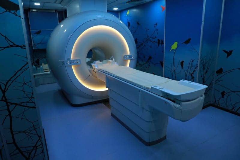
Recently upgraded system – 1st 1.5T digital MRI in Gujarat.
Relatively silent MRI (less noise)
Flared edges of tunnel – for less claustrophobia
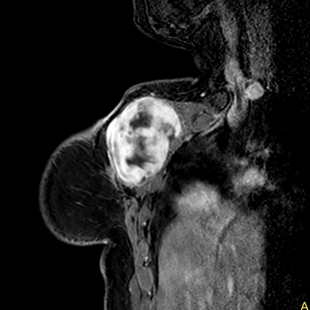
Dedicated coils for shoulder, knee, finger, TM joint.
Fast scan protocol for unstable patients.
Metal artifact reduction software for implant evaluation and surrounding soft tissues
3D spine software for spine evaluation in patient having scoliosis, kyphosis.
Non contrast perfusion in patients with renal failure for evaluating tumor perfusion.
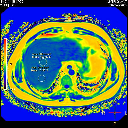
Liver fat quantification.
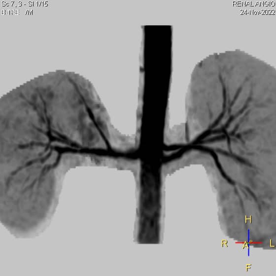
Non contrast angiography of aorta, renal arteries, upper limb, lower limb in renal failure patients whom contrast cannot be given.
Brain MRI – for stroke , tumor evaluation.
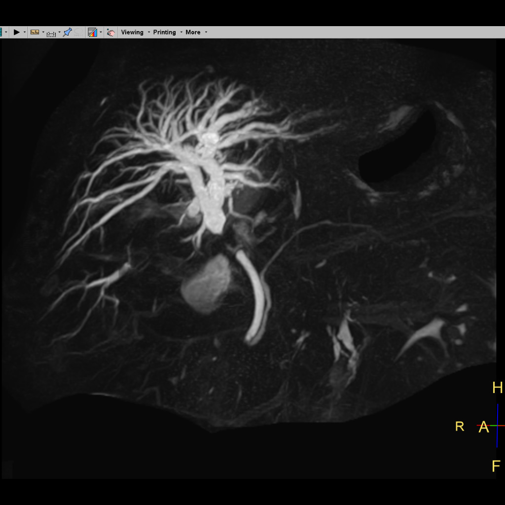
Brain angiography : non contrast / contrast angiography for narrowing in head and neck arteries.
Brain venography : non contrast / contrast venography for central venous sinus thrombosis.
PSP protocol : special protocol for parkinson’s plus syndrome with relevant calculations.
CT Paranasal sinuses : for diagnosing paranasal sinus diseases and to look for anatomical variations.
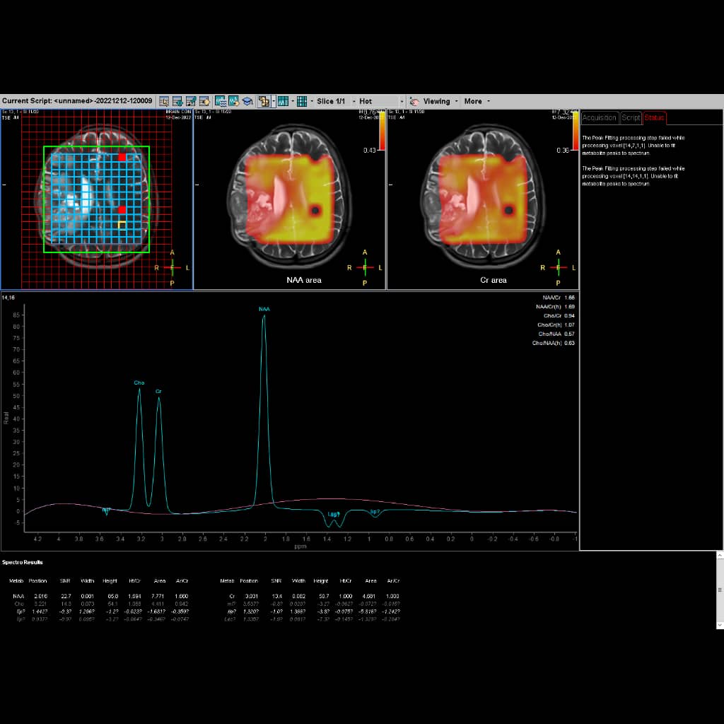
MRI spectroscopy : for suspicious cases not diagnosed on routine MRI.
MRI tongue : for pre-operative evaluation of tongue neoplasm.
TM joint evaluation : for internal disc derangement.
Temporal bone evaluation : for diagnosing recurrent cholesteatoma.
MRI sialography : for salivary duct evaluation
Spine : for disc prolapse, infection, tumors.
Musculoskeletal : for ligament, soft tissue injuries.
Perianal : for perianal abscess, sinus, fistula evaluation ; with diagrammatic representation
Defecography : for unresolved constipation, sphincter disorders, prolapse.
MRCP : non contrast evaluation for biliary and pancreatic ducts.
MRI liver triphasic – for liver tumors ; with MRI liver specific contrast agents
Multiparamagnetic MRI of prostate – for prostate neoplasm
Pelvis MRI – for endometriosis, ovarian / uterine / rectal neoplasm.